How to repair a damaged heart: key mechanism behind heart regeneration in zebrafish revealed
&width=710&height=710)
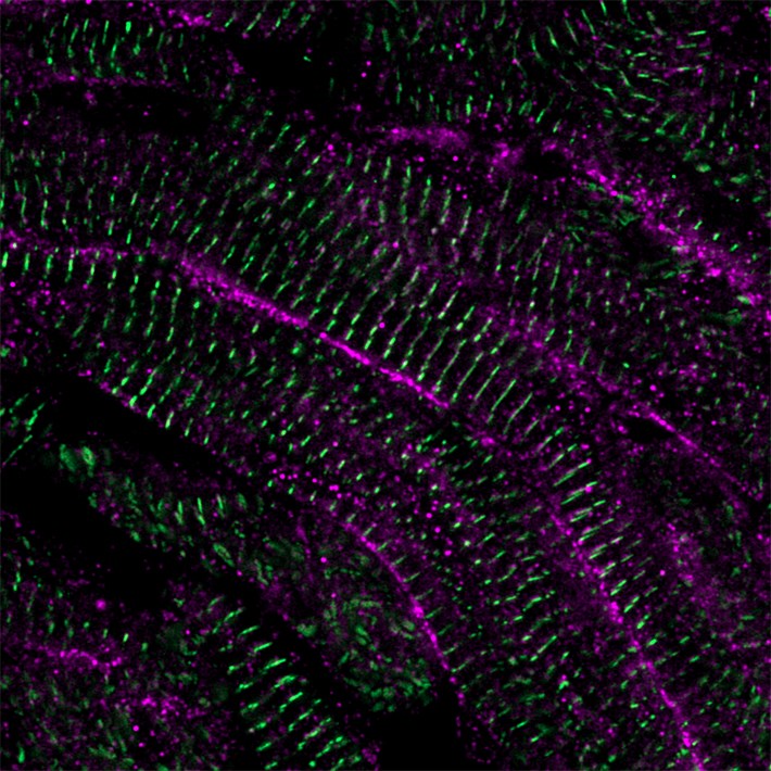
Figure 1: Credit: Phong Nguyen. Copyright: Hubrecht Institute.
Researchers from the group of Jeroen Bakkers (Hubrecht Institute) have used the zebrafish to shed light on their regenerative success. They discovered that newly formed heart muscle cells change their calcium handling over time and identified LRRC10 as a factor that drives these changes. LRRC10 thereby controls the decision between division and maturation of these cells. Researchers from the Leiden University Medical Center (LUMC) then showed that it had a very similar effect on human heart muscle cells and thus is evolutionarily conserved. The results of the study, published in Science on May 18th, show that examining the natural heart regeneration process in zebrafish and applying these discoveries to human heart muscle cells could contribute to the development of new therapies against cardiovascular diseases.
It is estimated that 18 million people die from cardiovascular diseases every year. Many of these deaths are related to heart attacks. In such an event, a blood clot prevents the supply of nutrients and oxygen to parts of the heart. As a result, the heart muscle cells in the obstructed part of the heart die, which eventually leads to heart failure. Although therapies exist that manage the symptoms, there is no treatment that is able to replace the lost tissue with functional, mature heart muscle cells and thereby cure the patients.
Zebrafish as a role model
Unlike humans, some species like zebrafish can regenerate their hearts. Within 90 days after damage, they fully restore their cardiac function. The surviving heart muscle cells are able to divide and produce more cells. This unique feature provides zebrafish hearts with a source of new tissue to replace the lost heart muscle cells. Previous studies successfully identified factors that could stimulate heart muscle cells to divide. Nevertheless, what happens to the newly formed heart muscle cells afterwards had not been studied before. Phong Nguyen, first author of the study, explains: “It is unclear how these cells stop dividing and mature enough so that can they contribute to normal heart function. We were puzzled by the fact that in zebrafish hearts, the newly formed tissue naturally matured and integrated into the existing heart tissue without any problems.”.
Calcium controls division and maturation
The researchers discovered that the cardiac dyad, a structure in the heart muscle cells regulating calcium movement, and one component specifically, called LRRC10, controlled division and maturation of heart muscle cells in the regenerating zebrafish heart. Nguyen: “Zebrafish heart muscle cells that lacked LRRC10 continued to divide during regeneration and remained immature. Excitingly, the opposite was also true. Induced expression of LRRC10 during regeneration prevented the division of the heart muscle cells”. Previous studies identified factors, such as stimulation of Nrg1/ErbB2 or inhibition of Hippo signaling, that drive uncontrolled cell division in the heart. This typically leads to the formation of enlarged hearts, called cardiomegaly. The researchers now found that LRRC10 prevented the formation of these ‘mega hearts’ as well, confirming the idea that it functions as a crucial factor determining division on the one hand and maturation on the other.
From fish to human
After Nguyen and his colleagues established the importance of LRRC10 in stopping cell division and initiating maturation of zebrafish heart muscle cells, they moved on to test if their findings could be translated to mammals. To this end, they collaborated with other institutes and induced the expression of LRRC10 in mouse and lab-grown human heart muscle cells. The human cells were derived from human pluripotent stem cells, generated by the LUMC hiPSC hotel. The evidence that LRRC10 is also important for maturation of human heart cells was obtained by LUMC researchers: postdoc Giulia Campostrini carried out the work under supervision of Milena Bellin and Christine Mummery. Bellin: “This work builds on our earlier studies into the maturation of cardiomyocytes: it was fantastic to see the role of LRRC10 being conserved between zebrafish and human”. Strikingly, LRRC10 changed the calcium handling, reduced cell division and increased the maturation of these cells in a similar manner as observed in zebrafish hearts. Nguyen: “It was exciting to see that the lessons learned from the zebrafish were translatable as this opens new possibilities for the use of LRRC10 in the context of new therapies for patients”.
Clinical impact
The study, published in Science, identified LRRC10 as a factor that has the potential to drive the maturation of heart muscle cells further through the control of their calcium handling. The lessons learned from the zebrafish heart could thereby have an important contribution to the development of new therapies. “The development of several therapies is currently hampered by the immaturity of heart muscle cells in culture. Therapies based on cell transplantation and in vitro drug testing platforms could for instance benefit from factors that are able to force the maturation. We now identified LRRC10 as one such factor,” says Jeroen Bakkers, last author of the study. LRRC10 could, by making cultured heart muscle cells more representative of the human situation, thus improve the chances of developing successful new treatments against cardiovascular diseases.
The study is the result of a collaboration between the Hubrecht Institute, LUMC, AMC, UMC Utrecht and Weizmann Institute. The study was financed by the Dutch Heart Foundation, Dutch CardioVascular Alliance and Stichting Hartekind, the Novo Nordisk Foundation Center for Stem Cell Medicine (reNEW) and the European Research Council (CoG Mini-HEART).