50 years of MRI: how the LUMC can make this indispensable technology affordable for the rest of the world
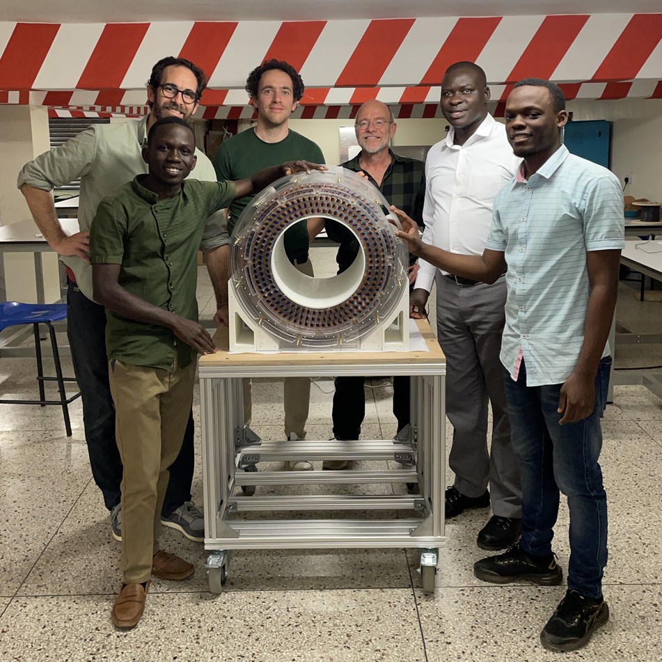
The LUMC team travelled to Uganda with their small MRI to show local engineers and students how to assemble and maintain the device.
&width=710&height=710)
A small and affordable MRI? Just replace expensive materials with cheap alternatives. “Unfortunately, it's not that simple,” says Andrew Webb, Professor of Radiology. “MRI is a complex technique. To make a smaller version, which can also be used in regions such as rural Africa, we had to design the system from scratch.
 The usual superconducting magnet was replaced by thousands of small magnets, and electrical components were redesigned to run on far lower power or even batteries. Algorithms that process MR images have also been modified so that they filter out noise from the outside. The result? A scanner weighing 75 kg, 50 by 50 centimetres in dimensions, which can be easily assembled without specialized tools. And material costs around 1% of the price of a normal MRI.
MRI for everyone
While in Europe there are as many as 35 MRI machines available per 1 million inhabitants, in Africa the number is less than one. “A machine like ours should enables doctors in Africa to diagnose and treat life-threatening diseases on the spot,” says Webb. Although the quality of a normal MRI cannot be matched, this small MRI also has added value for high-income countries. Webb: “It makes the scan more accessible. In theory, it could be placed in an ambulance to better help stroke patients, for example.”
Last year, the LUMC team shipped a system in kit-form to Uganda. There they trained local engineers and researchers to build and maintain the system themselves. “On a technical level, we have shown that we can make an affordable MRI, now we have to demonstrate that it can also be used in clinical practice.” In the coming months, the device will be used to train students from many other African countries.
Webb and colleagues have consciously chosen not to apply for a patent on their technique. “Our goal is to make MRI accessible to the whole world, and that will go faster if everyone has free access to our design.” The techniques developed by Webb and colleagues will be, or is already being, used in Paraguay, Spain and Uganda, where its application for the diagnosis of viral brain and musculoskeletal disorders is being investigated.
A historical perspective
“MRI has turned medicine and biomedical research upside down over the past 50 years,” says Mark van Buchem, head of the Radiology department. Until the 1970s, only X-rays were used in radiology. “That technique provided only limited information about the human interior. With the introduction of MRI, tissues could be assessed on the basis of many more characteristics. This resulted in a wealth of information with which much more accurate diagnoses could be made,” says Van Buchem. “It has become an indispensable device for patient care.”
Simple is not always easy
About 40 years ago, one of the first MRI scanners in the Netherlands was installed in the LUMC. To this day, the LUMC is still working on improving MRI techniques. Andrew Webb's group and others in the Gorter Centrum are also active in these areas. “On the one hand, we work on state-of-the-art technologies, while on the other hand we work on making things as simple and inexpensive as possible” says Webb. But that too is innovation. “Making things simple is extremely difficult,” he concludes.
In Nature, Webb, along with Johnes Obungoloch of Mbarara University of Science and Technology, describes five steps to make MRI scanners more affordable for the whole world.
&width=826&height=464)