Pulmonary & Musculoskeletal Imaging
This section is primary engaged in image processing for applications in Pulmonology and Musculoskeletal Imaging (PMI) in close collaboration with the corresponding clinical departments at the LUMC, including Orthopaedics and Rheumatology.
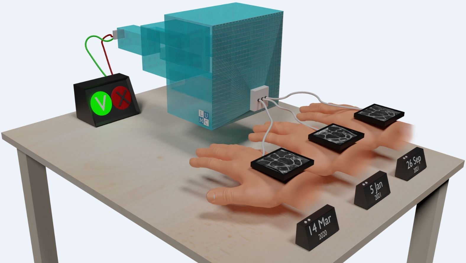
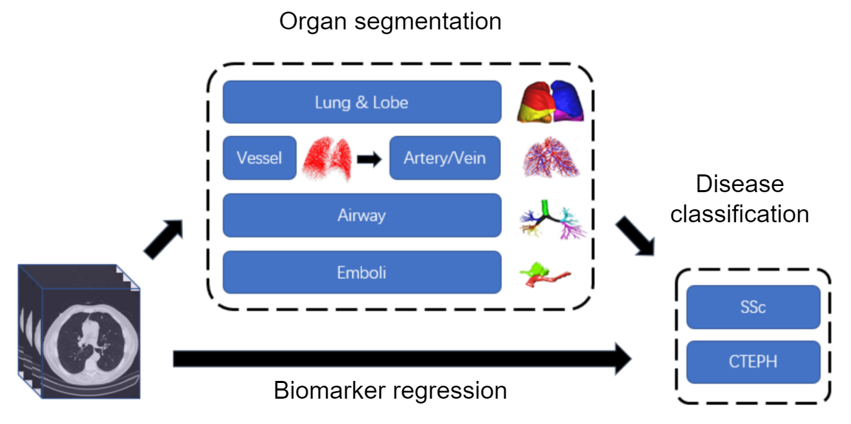
The pulmonary applications are focused on the quantification of lung diseases, such as pulmonary emphysema, interstitial lung disease and pulmonary embolism, using CT densitometry and automatic detection and assessment of vascular structures, as well as deep learning based scoring. The orthopaedic applications involve projects, in which the micro-motion of endoprostheses are measured and in which the optimal allograft is selected automatically for segmental bone reconstruction. Together with Rheumatology, our research is focused on deep learning based detection of early rheumatoid arthritis and its progression in MRI of the hands and feet.
Current projects:
Pulmonology
- Deep Learning-based Segmentation and Biomarker
- Deep Learning in Detecting Pulmonary Embolism in Computed Tomographic Pulmonary Angiography
Orthopaedics
- Automatic Structural Allograft Selection from Virtual Bone Bank in Segmental Reconstruction
Rheumatology
- AIMIRA: Artificial-Intelligence-based Comparative MR Imaging in Detecting Inflammatory Changes in Arthralgia Suspicious for Progression to RA (TTW-OTP)
Former projects
- Extremity MRI for Early Identification of Rheumatoid Arthritis (STW-HTSM)
- ESMIRA+: Deep Learning in the Detection of Early Inflammatory Signs in Rheumatoid Arthritis (CSC)
- Through the eyes of AI: safe and optimal integration of Artificial Intelligence in Radiology
- Evaluation of intra-operative CT and Navigation in Orthopaedic Oncology Surgery
- Detection and Quantification of the Pulmonary Vascular Tree (CSC)
- Automated Retinal Topographic Analysis with Magnetic Resonance Imaging
- PROTONS4vision: vision-sparing eye tumor treatment using proton therapy and MRI-only planning(TTW-HTSM)
- APPEAR: Quantification of Local Emphysema Progression
- Model-based Roentgen Stereophotogrammetric Analysis of Orthopaedic Implants
- Model-based Shape Matching of Orthopaedic Implants in RSA and Fluoroscopy
- Wear Measurements in Total Knee Arthroplasty (with Orthopaedic dept.)
- Fast Image Registration for Time-critical Medical Applications
- Investigation Towards Quantitative Measures from CT-images in Lung Diseases
- Demonstration of the Performance of a New Analytical Software Package Developed for Detection of Progress of Emphysema by CT (SPREAD)
- Software Development for the Detection and Assessment of Small Airways Disease in COPD With Multi Slice Computed Tomography
- Pollen Recognition in Microscopic Images
- Pollen Detection from Microscopic Images
- Labelling the Pulmonary Arterial Tree
- Improvement and Standardization of the Acquisition and Analysis of Angiographic Radiographs of Stenotic Renal Arteries
- Evaluation of Radiotherapy by Automatic Image Registration
- Measurement of Joint Space Width in assessing Osteoarthritis from Radiographs
- Detection and quantification of early ischemic changes
- Absolute Contrast Measurement in Digital X-ray Images
Our Team members
- Berend C. Stoel, team lead
- Els Bakker, senior researcher
- Kilany M.A. Hassan, senior researcher
- Y. Li, PhD student
- Jingnan Jia, PhD student
- Q. Du, PhD student
- Denis Shamonin, scientific programmer