Microscopic Image Analysis
Both of them are used as primary visualization modalities in biological research. At the same time, discovery potential of these data still largely remains underexploited as their analysis is currently limited to either manual or very basic automated methods. Our goal is to develop robust automated image analysis techniques for these modalities that would facilitate biological discovery.
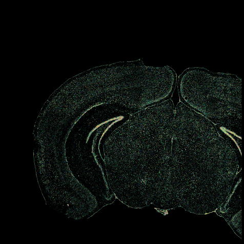
We perform fundamental and applied research in the area of biomedical image processing and analysis. We execute extensive validation studies of the developed techniques, both technically and clinically. We aim to impact the healthcare system by bringing research results close to the clinic, through collaboration with clinicians as well as with industry. While most of our research focuses on medical imaging, we are also interested in biological and genetic data. We have worked in particular on CT, MRI, IVUS and OCT imaging, in the brain, chest, heart, abdomen, vasculature and bones. In addition, we investigate highly heterogeneous data such as omics, imaging and mass-spectrometry combined, e.g. to produce new insights about structural and functional organization of the brain.
Current projects:
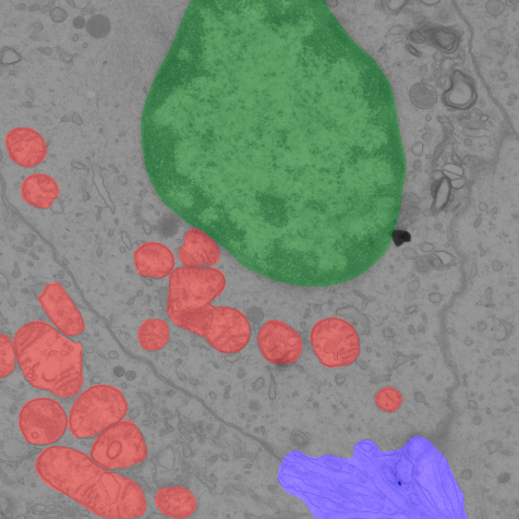
- Quantitative analysis of electron microscopy data
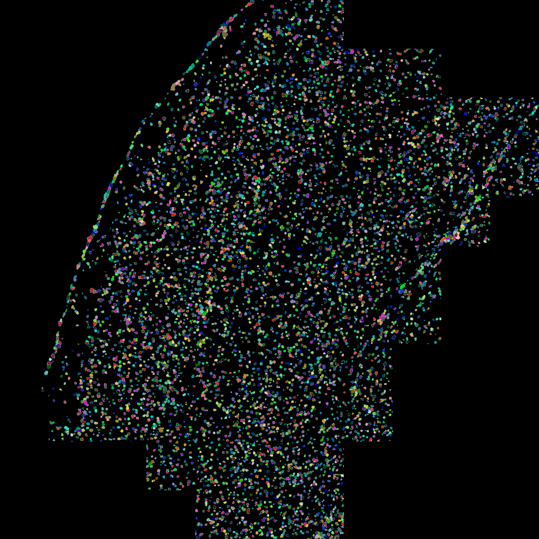
- DAPI cell segmentation for spatial transcriptomics data
Our Team members
- Oleh Dzyubachyk, Team lead